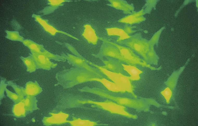
The fluorescence phenomenon has been observed for thousands of years and was mentioned in Chinese books as far back as 1500 BC. Several hundred years ago, Johann W. von Goethe described fluorescent light phenomenon in his Theory of Colors. He asked his readers to: “… dip a fresh piece of horse chestnut bark into a glass of water; the bark will immediately turn sky-blue. “ However, only today do we truly understand this phenomenon and have the ability to control and make use of its processes.
Applications & general facts
Fluorescence phenomenon
Fluorescence is a special form of luminescence, an optical phenomenon in cold bodies, in which a molecule absorbs a high-energy photon, and reemits this light at a lower-energy or longer-wavelength. The term fluorescence is named after the mineral calcium fluoride, which has been seen to exhibit this phenomenon.
The energy difference between the absorbed and emitted photons is released as molecular vibrations (heat). Usually the absorbed photon is in the ultraviolet part of the spectrum, and the emitted light (luminescence) is in the visible range, but this depends on the absorption curve and Stokes Shift of the particular fluorophore. The Stokes shift, so named after the Irish physicist George G. Stokes, is the difference (usually in frequency units) between the spectral positions of the band maxima (or the band origin) of the absorption and luminescence arising from the same electronic transition.
Generally, the luminescence that occurs at longer wavelengths than the absorption is stronger than those at shorter wavelength. The latter may be called an anti-Stokes shift. Each fluorescent material has its own unique value, basically a fingerprint that allows the material to be clearly identified.
Due to the fact that today‘s measurement technology is complex, expensive and only practical in labs, it has not always been possible to use fluorescence techniques for many applications. Lab-based measurement systems, while highly sensitive, are usually bulky and expensive.
The measurement of fluorescence demands complex, highly sensitive systems because the emission energy, the energy of the sample‘s fluorescence light, is nearly a million times smaller than the energy of the excitation. The fact that the emission has only a slightly different wavelength than the excitation (20-30 nm) is one of the challenges of this technique. In addition, lab-based measurement systems use a laser or high-pressure lamp (for variable wavelengths) to excite the sample and highly sensitive detectors, such as photomultipliers or ccd-detectors, to measure the emitted energy. The optical separation of excitation and emission is usually conducted at a measurement angle of 90°. This measurement principle is known as Off-Axis-Measurement and requires a very precise positioning of the excitation and emission beam on the sample. Therefore lab systems are very powerful and able to detect arbitrary fluorescent objects. However, these systems are very large, heavy, consume high amounts of energy and cannot be used under difficult environmental conditions due to the fact that temperature-differences, humidity or dirt particles have an enormous effect on the measured results. The operation of these systems normally requires trained personnel, which incurs additional costs. Thus the use of these systems is still limited to labs and research institutions.
Fluorescence has proven to be a versatile tool for myriads of applications. This powerful technique can be used to study molecular interactions in analytical chemistry, biochemistry, cell biology, physiology, nephrology, cardiology, photochemistry, and environmental science as well as in other areas. Fluorescence detection has three major advantages over other light-based investigation methods: high sensitivity, high speed, and safety. Safety here refers to the fact that samples are not affected or destroyed in the process and no hazardous byproducts are generated.
New developments in instrumentation, software, probes, and applications have resulted in a heightened popularity for a technique that was first observed over 150 years ago. There are many natural and synthetic compounds that exhibit fluorescence, and they have a number of different diagnostic, medical or biochemical applications.
For example, a fluorescent chemical substance or fluorophore can be attached to large biological molecule that is of interest and fluorescence can then be used to identify if the molecule of interest is present. This enables very specific analyses and, more importantly, quantitative results as the signal strength will depend on the number of fluorophores in the sample. Applications include DNA-sequencing, biochips, tumor and immunological marker detection.
Fluorescence is also used in many industrial applications, for example, to identify substances, surface coatings and final products. Applications include proof of authenticity (currency notes, expensive consumer products), inspection of coatings and tagging of fuel or oil. In addition, organic material when excited with UV-Light, will often display a strong auto-fluorescence and this attribute has a wide variety of applications without the need to use additional fluorescence markers. One such example would be the measurement of contamination (e.g. oil in water) and the detection of oil and fat residues on surfaces in hygiene testing.
Common applications
Immunofluorescence (IF): Immunofluorescence is used to detect the presence of antigens or antibodies. This technique involves applying specific antibodies that are labeled with a fluorescent dye to the sample under investigation. Using fluorescence microscopes, researchers can then visualize the localization and presence of these target molecules in cells or tissues.
Fluorescence in situ hybridization (FISH): FISH is a technique that uses fluorescently labeled DNA probes to detect specific DNA sequences in cells or tissues. This method is often used to identify chromosomal abnormalities, gene mutations, or to study microbial infections.
Fluorescence immunoassays (FIA): Fluorescence-labeled antibodies or antigens are used in immunoassays to determine the amount of a specific analyte in a sample. This method allows for sensitive and quantitative analysis of biomolecules such as proteins, hormones, or infection markers.
Fluorescence PCR (Polymerase Chain Reaction): Fluorescent dyes can be incorporated into PCR to monitor DNA amplification in real-time. This quantitative PCR (qPCR) enables rapid and accurate determination of the initial concentration of a DNA target sequence and is frequently used for diagnosing infections or genetic diseases.
Fluorescence cell analysis (Flow Cytometry): This method allows for the analysis and sorting of cells based on fluorescent labels. Fluorescent dyes are used to identify specific cell populations, examine the expression of surface molecules, or analyze intracellular components.
The use of fluorescence in in vitro diagnostics offers the advantage of high sensitivity, specificity, and multiplexing capability, as multiple fluorescent dyes can be detected simultaneously. This allows for complex and versatile analyses of biological samples in diagnostic research and clinical applications.
Explanation using PCR systems
Fluorescent dyes are used in Polymerase Chain Reaction (PCR) to monitor the progress of the amplification reaction in real time. This allows for a quantitative analysis of the initial concentration of the target DNA molecule in the sample. The advantage of fluorescence in PCR lies in the ability to track the amount of DNA produced during each amplification cycle in real time, rather than only at the end of the reaction.
Here is a simple example of using fluorescence in PCR:
Preparing the target DNA molecule: The DNA to be analyzed is mixed with specific primers (short DNA pieces that flank the region to be amplified) and a probe oligonucleotide marked with a fluorescent dye.
Performing PCR: PCR is initiated, during which the DNA is amplified in short cycles using DNA polymerase and the primers. During amplification, the fluorescent probe binds to the developing DNA chain.
Fluorescence detection: In each amplification cycle, the fluorescence is measured by a special detection system. As the amount of DNA increases, the fluorescence becomes more intense.
Real-time monitoring: The number of PCR cycles needed to reach a measurable fluorescence threshold (Ct value, threshold cycle) correlates with the initial concentration of the target DNA molecule in the sample. A lower Ct value indicates a higher initial concentration.
Advantages
Quantitative Analysis: Real-time PCR with fluorescence allows for the precise quantification of the initial concentration of the target DNA molecule.
Rapid Results: Real-time monitoring enables the quick generation of data, in contrast to conventional PCR where analysis only occurs after the reaction is complete.
Multiplexing: By using different fluorescent dyes, multiple target molecules can be simultaneously amplified and quantified in the same sample.
Early Detection: The method allows for the early detection of infections, genetic mutations, or other target molecules in diagnostic applications.
Therefore, real-time PCR with fluorescence is a powerful technique in molecular diagnostics and is used in numerous applications such as infection diagnostics, gene expression analysis, and the detection of genetic variations.

